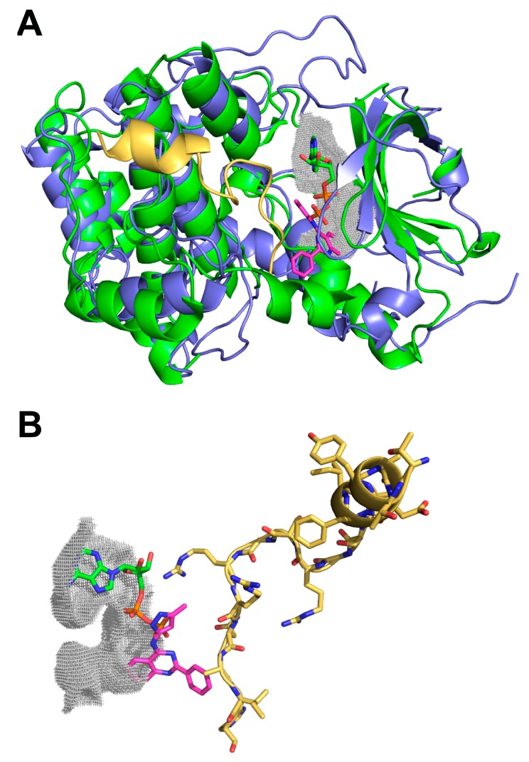
Figure 2.
Representation of PKI complexed to PKAC-alpha and the relative position of GSK-3 Inhibitor XIII in the ATP and substrate-binding sites of hNek2. (A) Structural alignment between PKAC-alpha (PDB: 1FMO) (blue) and hNek2 (PDB: 2W5A) (green), both displayed in cartoon representation, with a root mean square deviation of 2.010 Å. The PKI peptide inhibitor is depicted in yellow whereas the GSK-3 Inhibitor XIII compound is shown in magenta alongside the ADP molecule (green) inside the ATP-binding site; (B) Detailed representation of the most favorable conformation adopted by GSK-3 Inhibitor XIII (magenta) that could block the access to the substrate-binding site.

