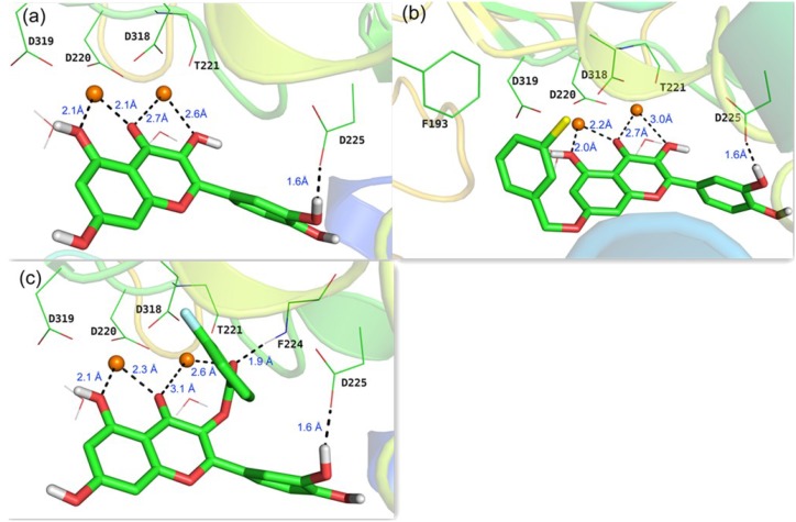
Figure 3.
Predicted binding modes in the active site of a reported X-ray structure of HCV RdRp (PDB code: 1GX6 [22]) from docking studies: (a) quercetin 2; (b) compound 3i; (c) compound 4f. The two Mg2+ ions are indicated as orange spheres, carbon atoms are shown in green, oxygen in red, chlorine in yellow, and fluorine in blue.

