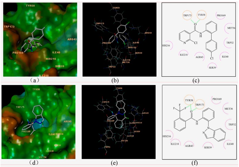
Figure 4.
Surflex-Docking of boscalid to complex II. (a) Connolly surface of complex II with boscalid shown as a stick model. (b) Interaction of boscalid and amino acid residues near the ligands (3D diagram). (c) Interaction of boscalid and amino acid residues near the ligands (2D diagram). (d) Connolly surface of complex II with compounds 3c shown as a stick model. (e) Interaction of compounds 3c and amino acid residues near the ligands (3D diagram). (f) Interaction of compounds 3c and amino acid residues near the ligands (2D diagram). The orange dotted line circles show the amino acids that participated in hydrogen bonding. The magenta dotted semicircle show the amino acids that participated in the van der Waals interactions. The hydrogen bond interactions are shown as green dotted lines.

