Figure 4.
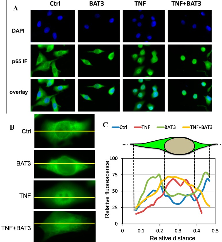
BAT3 does not inhibit nuclear NF-κB translocation. (A) A549 cells grown on coverslips were incubated with 25 µM BAT3 for 2 h and then stimulated with 2000 IU/mL TNF for 30 min. After cell fixation, nuclei were visualized by blue DAPI staining and nuclear translocation of NF-κB was monitored by overlay of blue DAPI staining with anti-p65 green immunofluorescence microscopy; (B) Close-ups of NF-κB p65 immunofluorescence intensities across cytoplasmic and nuclear cell compartments for the difffeent treatments; (C) Cross-cellular (yellow line) fluorescence intensity plots were determined by Image J software (National Institutes of Health, Bethesda, MD, USA, version 1.49c, 2014) for the different treatments. For control and BAT3 treated cells, p65 immunofluorescence can be observed predominantly in the cytoplasm, with a relative lower fluorescence intensity across the nuclear region as expected. However, upon evaluating immunofluorescence signals following TNF or combined TNF + BAT3 treatment, p65 signal intensity is shifted more exclusively towards the nucleus as compared to the intensities observed in the cytoplasmic periphery.

