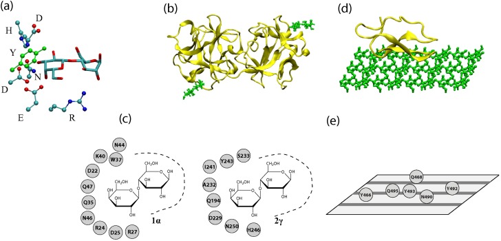
Figure 1.
(a) A three-dimensional representation of a binding pocket of a lectin (2γ site of the ricin B chain). Only the sugar residues and amino acid residues involved in ring-stacking and hydrogen bonding are shown. Hydrogen and backbone atoms are excluded for clarity. (b) A three-dimensional representation of a lectin (ricin chain B, PDB Code 2AAI) with lactoses bound at sites 1α (lower-left) and 2γ (upper-right). (c) A depiction of binding sites 1α and 2γ with possible hydrogen bond and ring stacking residues shown. (d) A three-dimensional representation of the cellulose binding module (CBM) of a CAZyme (Cel7A with cellulose modeled by M. Crowley’s group) (e) Residues of Cel7A’s CBM that interact with cellulose. Cellulose chains are represented by gray stripes on the plane.

