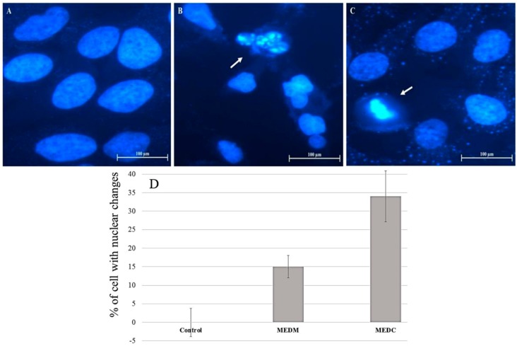
Figure 4.
Morphological changes of HeLa cells after treatment with ME for 24 h followed by DAPI staining. (A) Fluorescence microscope photographs of untreated cells; (B) cells treated with 0.2 mg/mL MEDM and (C) cells treated with 0.2 mg/mL MEDC; (D) DAPI staining quantification. Arrows indicate apoptotic bodies of nuclear fragmentation and chromatin condensation (B and C, respectively). Magnification ×400.

