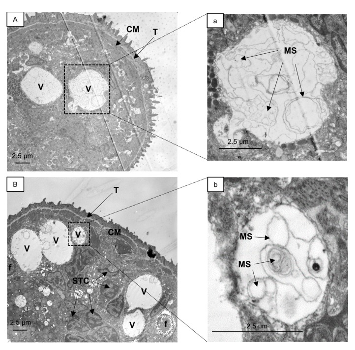
Figure 2.
Transmission electron micrographs of S. mansoni schistosomula exposed for 24 h to 5 µM 27 (A) or 23 (B). Enlargements of marked areas in (A,B) are depicted in (a,b), respectively. A fusion event between vacuoles is pictured in (B). Vacuoles with internal membranes (a,b) and onion-like multi-lamellar structures are visible (b). CM, circular muscle; f, flame cell; MS, multi-lamellar structures; STC, nucleus of subtegumental cell; T, tegument; V, vacuoles.

