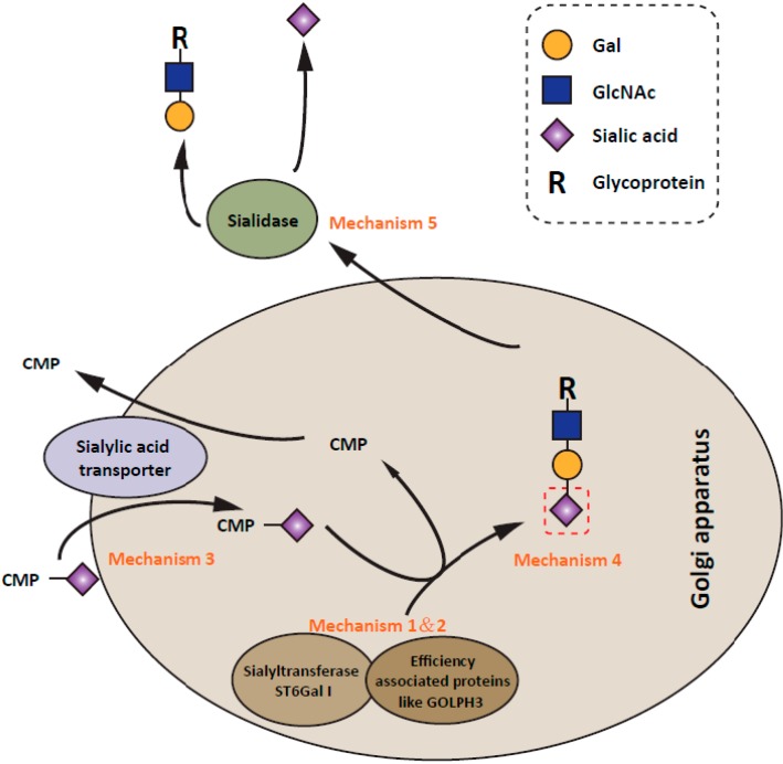
Figure 2.
Schematic representation of the regulation of α2,6 sialylation expression. The expression level of surface α2,6 sialylation is increased in tumors by several mechanisms. (1) Most commonly, ST6Gal I transcription is regulated by some transcription factors and methylation modification [18,31,32,33,34,35,36,37,101,130]; (2) Some factors like GOLPH3, recently, has been shown to interact with ST6Gal I, thereby modulating the efficiency of the sialylation [73]. (3) In addition, the expression levels of sialic acid transporter could affect α2,6 sialylation expression by regulating the donor substrate reservoir of ST6Gal I [118,119]; (4) The distribution as well as the amount of α2,6 sialic acids on cell surface also depend on the expression pattern of oligosaccharide acceptors [127,128,129]; (5) Further, as observed in many types of tumors, down-regulation of sialidases is usually accompanied by the upregulation of α2,6 sialylation expression [120,121,122,123,124].

