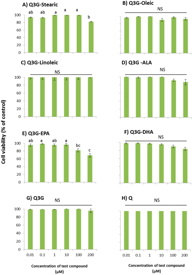
Figure 4.
Dose-dependent cytotoxicity of fatty acid esters of Q3G in WI-38 cells; (A) Stearic acid ester of Q3G; (B) Oleic acid ester of Q3G; (C) Linoleic acid ester of Q3G; (D) ALA ester of Q3G; (E) EPA ester of Q3G; (F) DHA ester of Q3G; (G) Q3G and (H) Quercetin. Cells were pre-incubated with test compounds for 48 h. Cell viability was presented as percentage related to the control. Control contains cells with no incubation of test compounds. Data are expressed as mean ± SEM (n = 6), means sharing the same letter within test compound are not significantly different (p ≤ 0.05).

