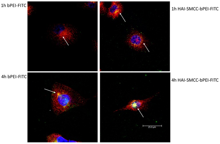
Figure 6.
Co-localization of transferrin (Tf) and bPEI or HAI-SMCC-bPEI polyplexes in H1299 was imaged by the confocal microscope. H1299 cells were incubated with Tf fluorescently labeled with Texas Red (Tf-Texas Red) and bPEI or HAI-SMCC-bPEI polyplexes prepared with 50 pmol of siRNA at N/P = 5 for 1 and 4 h. The polymers were fluorescently labeled with FITC and shown in green. Tf-Texas Red was shown in red, and DRAQ5 stained nuclei were shown in blue. The arrow pointing to the yellow dots represents co-localization spots of polyplexes and Tf.

