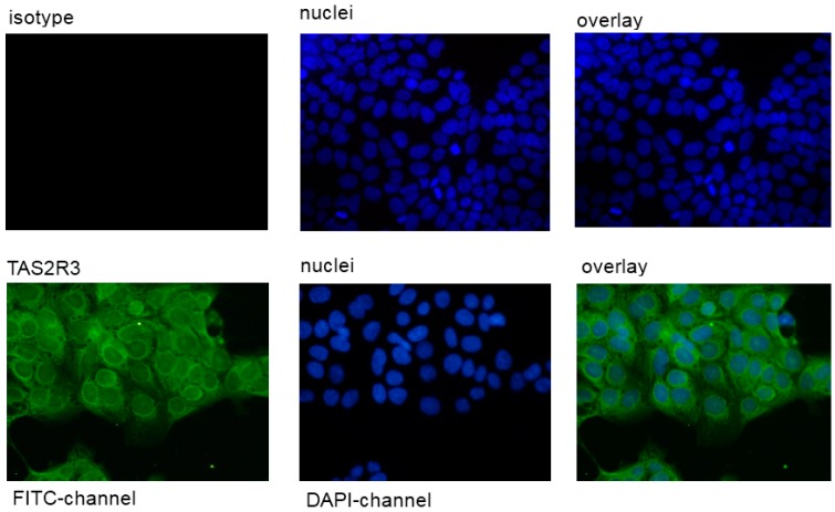
Figure 2.
Staining of the human cell line JEG-3 against TAS2R38. The rabbit anti-TAS2R38 antibody was visualized by a secondary FITC-coupled antibody in the FITC channel. The isotype control showed no fluorescence signal in this cell line. The pictures were photographed at magnification of 400×. The bar corresponds to 50 µm.

