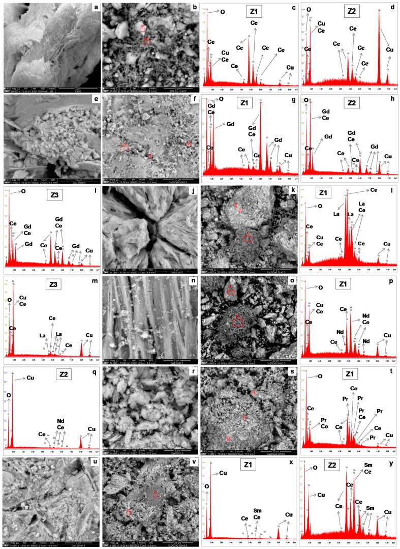
Figure 3.
SEM images of CeO2 (a); Cu/CeO2 (b) and respective EDS spectra of zones marked as Z1 (c) and Z2 (d); Ce-Gd (e); Cu/Ce-Gd (f) and respective EDS spectra of zones marked as Z1 (g), Z2 (h) and Z3 (i); Ce-La (j); Cu/Ce-La (k) and respective EDS spectra of zones marked as Z1 (l) and Z3 (m); Ce-Nd (n); Cu/Ce-Nd (o) and respective EDS spectra of zones marked as Z1 (p) and Z2 (q); Ce-Pr (r); Cu/Ce-Pr (s) and respective EDS spectra of zone marked as Z1 (t); Ce-Sm (u); Cu/Ce-Sm (v) and respective EDS spectra of zones marked as Z1 (x) and Z2 (y).

