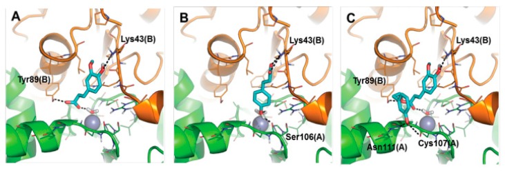
Figure 25.
Predicted binding mode of 2 (panel A), 3 (panel B) and 8 (panel C) towards the active site of Rv3588 β-CA from Mtb. Monomer A is colored green, monomer B is colored orange. Small molecules are shown as cyan sticks. Residues within 5 Å from the Zn(II) ion and those in contact with the inhibitors are shown as lines. H-bond interactions are highlighted by black dashed lines. Residues involved in H-bonds are labeled. The catalytic Zn(II) ion is shown as a grey sphere [85].

