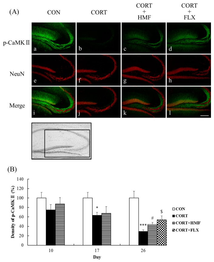
Figure 6.
Effects of HMF on the expression of phosphorylated CaMK II and NeuN immunoreactivity in the corticosterone-induced depressive mouse hippocampus. (A) Sagittal sections on Day 26 after continuous corticosterone injections were stained with specific antibodies, either anti-phospho-CaMK II (green; a, b, c, d) or anti-NeuN (red; e, f, g, h). Each signal was merged in i, j, k and l, respectively. The scale bar shows 200 µm. The location of the captured images in the hippocampus and quantification is shown with a square (1.0 mm2). (B) A quantitative analysis of phospho-CaMK II-positive signal density using ImageJ software. Values are means ± SEM (Day 10; n = 4, Day 17; n = 8, Day 26; n = 8–10). Symbols show significant differences between the following conditions: CON vs. CORT (* p < 0.05, *** p < 0.001), CORT vs. CORT + HMF (# p < 0.05), and CORT vs. CORT + FLX ($ p < 0.05).

