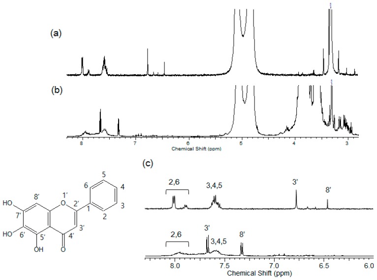
Figure 4.
(a) 1H-NMR spectrum of baicalein (Solvent: D2O:MeOD, 70:30, v/v); (b) 1H-NMR spectrum of cysteinyl β-CD/baicalein complex (Solvent: D2O:MeOD, 70:30, v/v); (c) Enlarged spectrum of the aromatic region (top: baicalein, bottom: cysteinyl β-CD/baicalein complex). The inset shows the chemical structure of baicalein.

