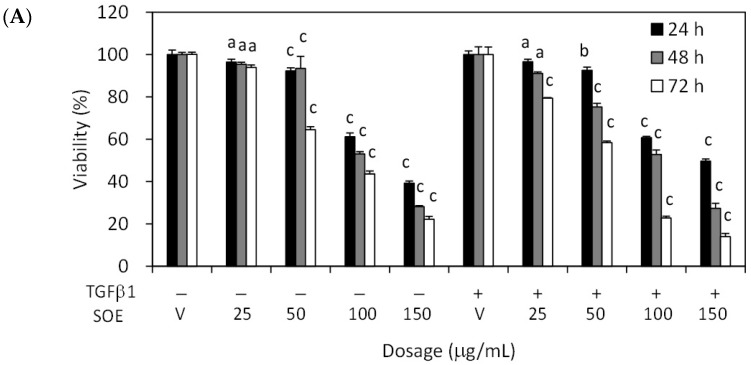
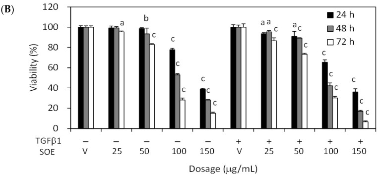
Figure 2.
Effects of concentration and duration of SOE treatment on proliferation of endometrial cancer cells, with or without TGFβ1 (2.5 ng/mL) stimulation. (A) HEC-1A cells and (B) RL95-2 cells. Cancer cells were cultured in 96-well plates at a density of 1 × 10⁴ cells/well. Cells were treated with 0–150 μg/mL SOE for 24–72 h. Cell viability was measured using 3-(4,5-dimethylthiazol-2-yl)-2,5-diphenyltetrazolium bromide (MTT) assay. Three independent experiments were conducted; results are expressed as mean ± SD. A significant difference from the respective vehicle, according to Student’s t-test, was indicated as “a” for p < 0.05; “b” for p < 0.01, and “c” for p < 0.001.


