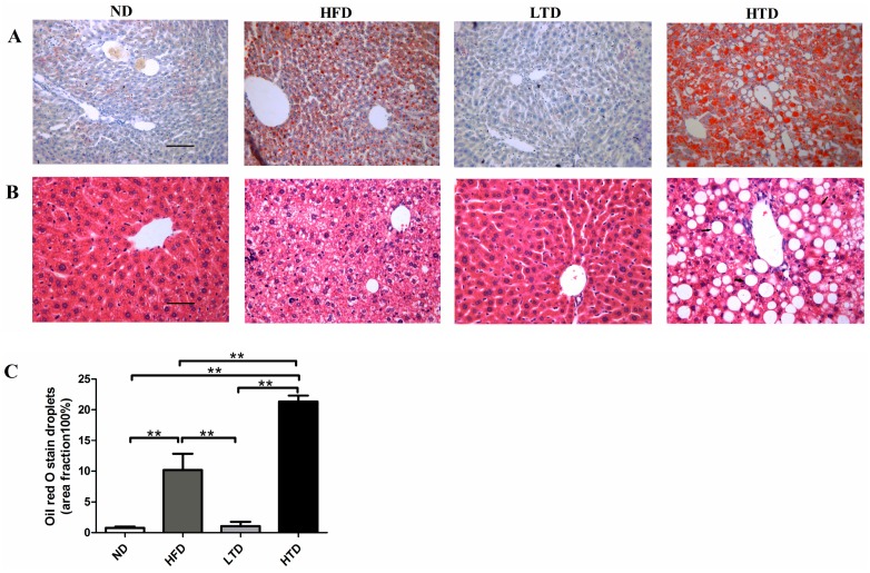
Figure 4.
Hepatic histology. (A) Oil-Red-O staining of liver sections in ND, HFD, TD and HTD groups post 20 weeks diet. Micrograph exhibiting oil droplets levels in the liver (magnification: 200× g) and red droplets in the picture exhibiting lipid droplets; (B) H&E staining of liver sections in ND, HFD, TD and HTD groups post 20 weeks diet (magnification: 200×) and white vacuoles representing lipid droplets. Scar bars indicate 100 μm; (C) Quantitative analysis of oil droplets, **: p < 0.001. Data are shown as mean ± SD.

