Figure 6.
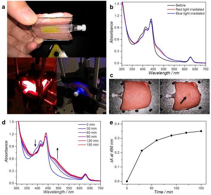
Irradiation of L134 liposomes through a thick slice of pork fillet. (a) Photographs of the experimental setup used for red and blue light irradiation. The 2 mm cuvette holding 400 µL L134 ([DLPC] = 10 mM) is covered with 7 mm pork fillet and irradiated for 2 h from above with a collimated 110 mW 630 or 450 nm beam (3 mm spot size, 1.6 W/cm2 intensity, 11.2 kJ/cm2 light dose); (b) UV-vis absorbance spectra of the sample before (black) and after irradiation with 450 nm (blue) or 630 nm light (red); (c) Photographs of the pork fillet after red (left) or blue light irradiation (right). Upon blue light irradiation, a clear “burn mark” was observed, as indicated with the arrow. UV-vis absorbance spectra (d); and the absorbance difference at 490 nm (e) as a function of irradiation time for L134 liposomes irradiated through 7 mm pork fillet with 300 mW 630 nm light (4.2 W/cm2). Experiment performed once. Arrows indicate the evolution of the spectrum in time near the indicated wavelengths.

