Figure 3.
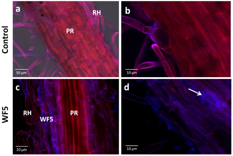
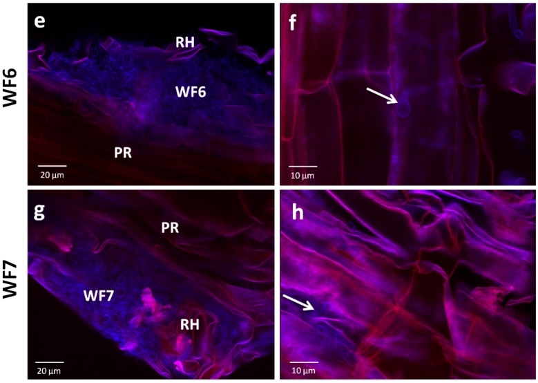
Test for the ability of the putative fungal endophytes to colonize finger millet roots using confocal scanning laser microscopy. (a–b) Representative pictures of root tissues inoculated with the buffer control; (c–h) Representative pictures of root tissues inoculated with each putative fungal endophyte as indicated. Fungi fluoresce purple-blue due to staining with calcofluor. Plant tissues appear red due to auto-fluorescence. White arrows point to fungi inside the tissues. Abbreviations: PR, primary root; RH, root hair.


