Figure 1.
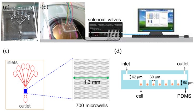
Automatic microfluidic platform for calcium imaging. (a) Photograph of the microfluidic chip (scale bar = 1 mm); (b) Experimental setup of the automated platform with the microfluidic chip and solenoid valves (scale bar = 2 mm); (c) The microfluidic chip contains 700 microwells that are 30 μm in diameter to array cells and six inlets to perfuse different solutions in a single experiment; (d) A cross-section schematic of the microfluidic device.

