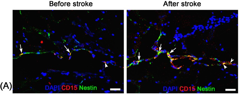
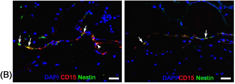
Figure 5.
Immunofluorescence studies of the activation of endogenous NSCs. Representative anti-CD15 (red) and anti-Nestin (green) immunofluorescence staining micrographs show the presence of CD15+Nestin− and CD15+Nestin+ cells in the SVZ (arrows) and beginning of RMS (arrowheads) before the establishment of stroke both in pure stroke mouse (A) and Ara-C-treated mouse (B). There are abundant CD15+Nestin− and CD15+Nestin+ cells in the SVZ and RMS in pure stroke animals eight days after stroke. Almost no CD15+Nestin− or CD15+Nestin+ cells are present in stroke animals after administration of Ara-C for seven days. Bars = 25 μm.


