Figure 2.
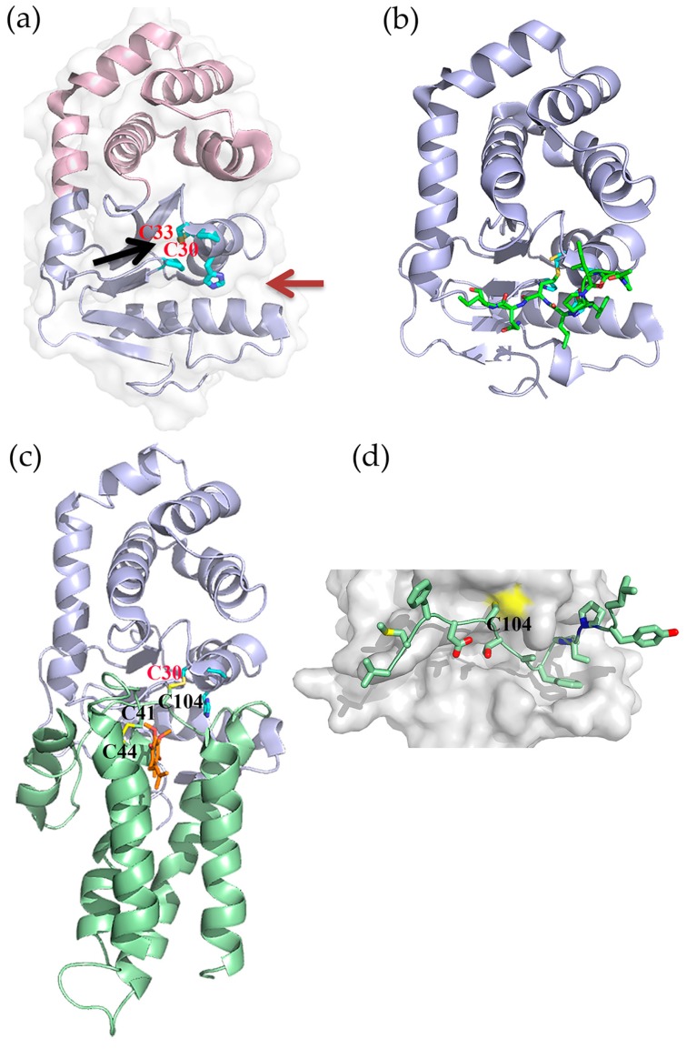
(a) Cartoon representation of EcDsbA (PDB 1FVK); thioredoxin fold shown in light blue and helical insert in light pink. Red and black arrows indicate the hydrophobic groove and hydrophobic patch, respectively; (b) Substrate peptide binding surface of EcDsbA (PDB 3DKS). Peptide and enzyme are shown in green and light blue respectively; (c) Crystal Structure of the EcDsbA–EcDsbB–UQ complex (PDB 2HI7). EcDsbA and EcDsbB are shown in cartoon representation (light blue and green respectively). DsbA Cys30 and DsbB Cys41,44, and 104 are displayed in stick representation. UQ molecule bound to DsbB is displayed in stick representation (orange); (d) Close-up view of the DsbB loop interaction site with the hydrophobic groove of EcDsbA. The DsbA the active site residues (Cys30-Pro-His-Cys33) and cis-proline residue are displayed in stick representation (cyan) in panels (a) to (c).

