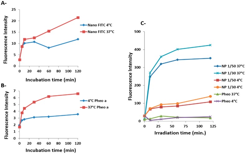
Figure 3.
Evaluation of cellular uptake of Pheo-a by flow cytometry versus incubation time and temperature. (A) {PEO(2000)-b-PCL(2600)} copolymer micelles labeled with fluorescein; red line, T = 37 °C; blue line, T = 4 °C; (B) Free Pheo-a (10−6 M; red line, T = 37 °C; blue line, T = 4 °C; (C) Loaded Pheo-a in copolymer micelles at molar ratios of 1/50 and 1/30. Each set of experiments and experimental conditions were performed twice.

