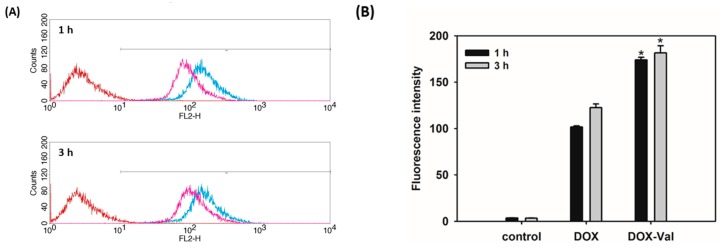
Figure 2.
Cellular accumulation efficiency of DOX-Val evaluated by flow cytometry in MCF-7 cells. DOX and DOX-Val (10 μM) were incubated for 1 h and 3 h in MCF-7 cells. (A) Histograms of all experimental groups, control (red), DOX (pink), and DOX-Val (blue), are shown; (B) The mean value of the fluorescence intensity of each group is presented. Data are presented as means ± standard deviation (n = 3). * p < 0.05, compared with DOX group.

