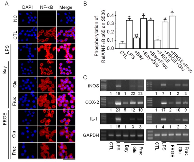
Figure 4.
Sugars block the anti-inflammatory effect of FRGE in LPS-stimulated RAW264.7 cells through NF-κB activation. (A) Nuclear translocation of NF-κB upon treatment with FRGE and either glucose or fructose. RAW264.7 cells were pretreated with Bay 11-7085, FRGE, and/or glucose or fructose for 12 h and then exposed to LPS for 1 h. Fluorescent cells are labeled with the NF-κB-specific antibody and Cy3-conjugated anti-rabbit IgG. The nucleus was stained with DAPI. The primary antibody was omitted in the negative control (NC). No red fluorescence was observed in the NC; only the DAPI stain was observed (blue color). The scale bar represents 50 μm; (B) FRGE-induced NF-κB suppression was decreased by co-treatment with glucose or sucrose in LPS-stimulated RAW264.7 cells. Total and phosphorylated NF-κB p65 levels were analyzed by ELISA using total cell lysate. Each bar represents the mean ± SD of three independent experiments. The plus (+) sign represents conditions with treatment. * p < 0.05 compared with the control (CTL). † p < 0.05 compared with the LPS treatment; (C) FRGE-induce reduction in the expression of iNOS, COX, and IL-1 mRNA was decreased by treatment with glucose or fructose in LPS-activated RAW264.7 cells. Total cell lysates were obtained using lysis buffer and subjected to RT-PCR. First-strand cDNAs were synthesized from 1 μg of total RNA isolated from the RAW264.7 cells, and the same concentration was used as the template for PCR. GAPDH was used as a loading control for the mRNA expression levels. The numbers below the PCR band represent the normalized ratio of the expression levels of iNOS, COX-2, and IL-1 to those of GAPDH for each lane. Bay, Bay 11-7085; Glu, glucose; Fruc, fructose.

