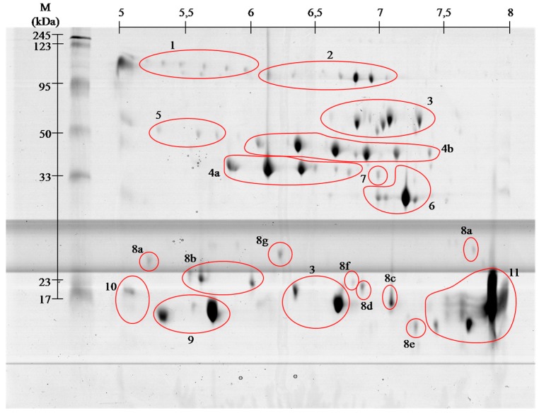
Figure 2.
Representative 2-D protein map in 5–8 pH range, obtained from V. berus berus venom with identified protein groups shown: 1, Angiotensin-like peptide; 2, Metalloproteinase H3; 3, l-amino acid oxidase; 4, Serine proteases: (a) VLSp and (b) nikobin; 5, Beta-fibrogenase brevinase; 6, Cysteine rich venom protein; 7, Snake venom metalloproteinases; 8, Snaclec: (a) rhinocetin, (b) snaclec 14, (c) snaclec B6, (d) echicetin, (e) snaclec 1, (f) rhodocetin/A13, and (g) jerdonibitin; 9, Acidic phospholipases; 10, Basic phospholipases; and 11, Neutral phospholipase. The proteins were separated by isoelectrofocusing at pH range 3–10, then distributed on polyacrylamide gels by SDS-PAGE and stained with colloidal Coomassie Brilliant Blue G-250. Molecular weight (MW) and pH 3–10 scale are shown.

