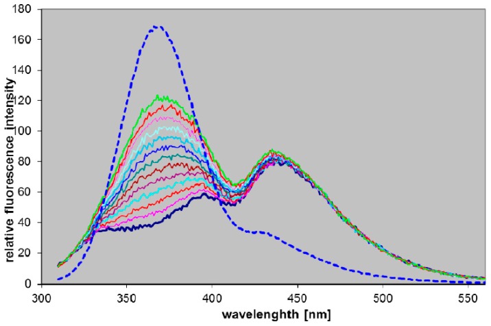
Figure 3.
Fluorescence changes during the incubation of 16 µM N7-ribosyl-8-azaDaPur in 1000-diluted blood in the presence of 25 mM phosphate. Spectra, excited at 300 nm, were recorded every 5 min, and after 60 min an aliquot of the purified calf PNP was added, and fluorescence recorded after next 3 min (dotted line, 5-fold diminished relative to remaining curves). Note that the first (lowest) curve reflects blood fluorescence background, and minimum at 415 nm is due to the re-absorption by hemoglobin.

