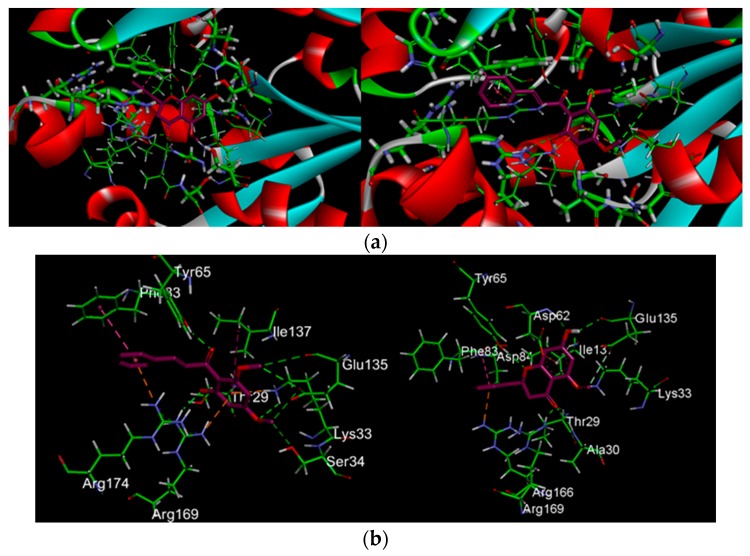
Figure 3.
(a) Interactions of ligands with UCK2 protein as identified by in silico docking analysis; (b) Ligands interacting with the amino acids residues in the active sites of UCK2 protein; (c) Surface representation of the hydrophobic contacts of bound ligand and the ligand binding pocket of UCK2 protein shown as translucent blue surface. Ligands shown as a ball-and-stick model with purple indicating carbon atoms, white for hydrogen, and red for oxygen Hydrogen bonds shown in green dotted lines, electrostatic in yellow, and hydrophobic in purple.


