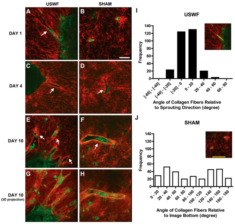
Figure 8.
Intrinsic cellular autofluorescence in second-harmonic generation microscopy of endothelial cell bands (green) suspended in unpolymerized collagen type-I fibers (red) on (A,B) day 1; (C,D) day 4; and (E,F) day 10 incubation treated by ultrasound standing wave field (USWF) or sham-exposure, arrows show endothelial cell sprouts in scale bar of 50 μm, and the histograms of the occurrence frequency of collagen fiber angles in (I) USWF- and (J) sham-exposed constructs. collagen gels, courtesy of [76].

