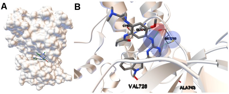
Figure 2.
(A) the tri-dimensional structure of the human wild-type EGFR kinase domain for the instance 4zau in which the cleft can be easily observable. The molecular surface is represented using ADTools (version 1.5.6, The Scripps Research Institute, California, EEUU). The computed (in green) and the co-crystallized ligands are also represented. The low value of the RMSD of the computed ligand returned by the algorithm MOEA/D does not allow the two ligands to be represented separately given that they overlap; (B) the molecular interactions between the ligand AZD9291 and the EGFR domain kinase. The H-bonds are represented by green spheres. The atoms involved in these H-bonds are represented by colored spheres according to the atom type. In this case, the nitrogen and oxygen atoms are represented in blue and red spheres, respectively. The aminoacids closer to the interactions and the aminoacid(s) involved in the H-bond(s) are also labeled.

