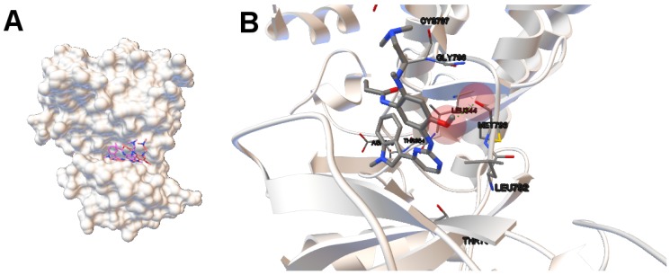
Figure 4.
(A) the tri-dimensional structure of the human wild-type EGFR domain kinase. The molecular surface is represented using ADTools. The computed and reference ligands are represented in pink and red, respectively. Both ligands’ conformations are bound on the outer edge of the cleft of the ATP-binding; image (B) shows the molecular interactions between the computed conformation of the ligand AZD9291 and the EGFR domain kinase (its secondary structure is also shown). The atoms involved in the H-bonds are represented by colored spheres. The H-bond is represented by green spheres.

