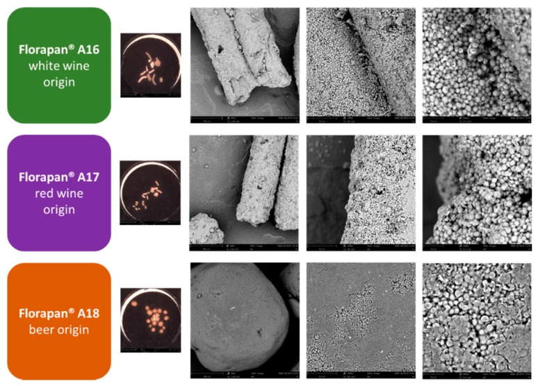
Figure 1.
The SEM images of the active dry yeast surface for the commercial preparations Florapan® A16 (A16), Florapan® A17 (A17), and Florapan® A18 (A18). Each preparation was visualized with an optical camera (~20X) (small colored image on the left) and with scanning electron microscope (SEM) (~250X, ~1500X, ~5000X) (grey images).

