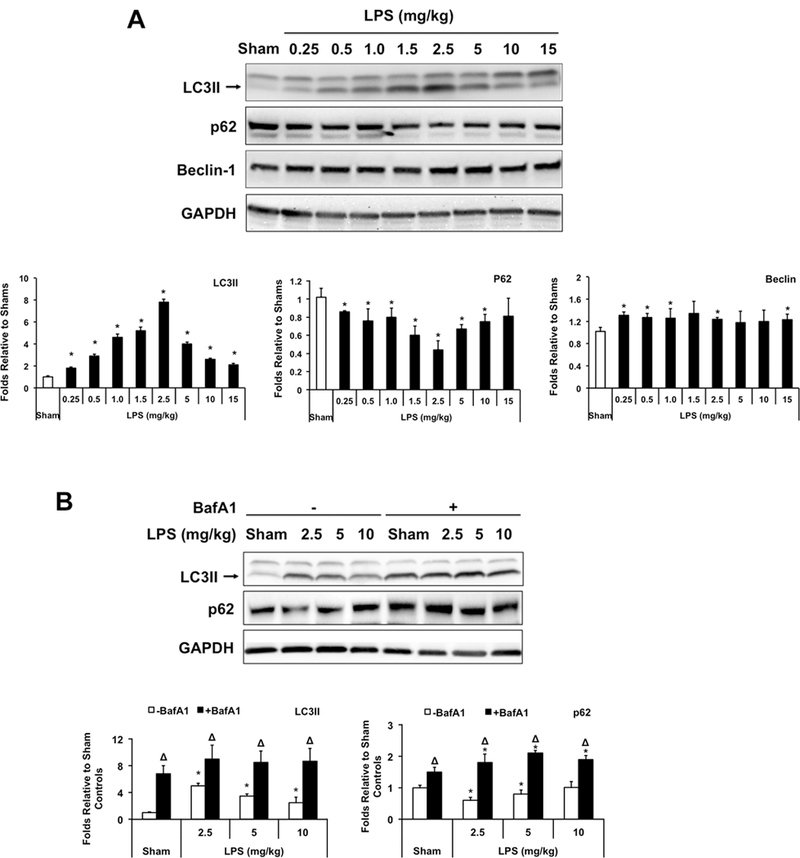
Figure 1.

Changes of cardiac autophagy in response to different doses of LPS. Wild-type C57BL/6 mice were given LPS via i.p. at indicated doses, heart tissues were harvested 18 hours later and total tissue lysates were prepared. A. Levels of LC3II, p62, Beclin and GAPDH were analyzed by Western blots using GAPDH as a loading control. B. LPS-induced autophagy flux was further confirmed by comparison of LC3II and p62 between animals with and without treatment of bafilomycin A1 (BafA1, 1.5 mg/kg). Results were quantified by densitometry and expressed as fold changes relative to shams. All values are means ± SEM. Significant differences are shown as * for sham vs. LPS-treated and Δ for vehicle vs. BafA1-treated groups (p < 0.05, n = 5, Mann-Whitney U test).
