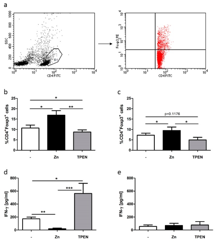
Figure 2.
Zinc deficiency and zinc supplementation adversely influenced Treg differentiation in MLCs. Here, 2 × 10⁶ PBMCs/mL remained untreated or were pre-incubated with 50 µM zinc (black bars) or 1.5 µM N,N,N′,N′-Tetrakis(2-pyridylmethyl)ethylenediamine (TPEN) (grey bars) for 15 min. MLCs were generated for five days. (a) One representative dot blot of gated (polygon, R1) viable CD4+ activated T cell blasts is displayed showing side scatter (SSC) and CD4-FITC (fluoresceinisothiocyanat) staining. Gating procedure was established in reference [16]. (b) Only cells of R1 (red dots) are displayed and were analyzed regarding CD4-FITC and FOXP3-PE (phycoerythrin) staining. These Tregs were calculated using FACS analysis in MLCs (activated T cells) and (c) PBMCs (resting T cells). The concentration of the pro-inflammatory cytokine interferon (IFN)-γ was measured in (d) MLCs and (e) PBMCs. All data are shown as means + SEM of n = 6 independent experiments (* p < 0.05, ** p < 0.01, *** p < 0.001, Student’s t-test).

