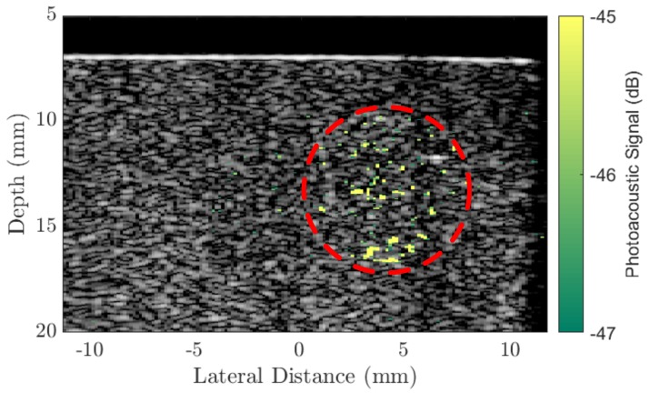
Figure 2.
A conventional B-mode plane-wave image (9 compounding angles) of a tissue-mimicking agar phantom with an overlaid photoacoustic image generated by a 8 mm diameter inclusion of Au50 AuNRs (concentration 20 μg/mL) at a depth of 3mm below the surface and a laser fluence 2 mJ/cm2 at the surface. The PA signal is referenced to the maximum of the B-mode image (dB). Red circle indicates the region of AuNRs.

