Figure 2.
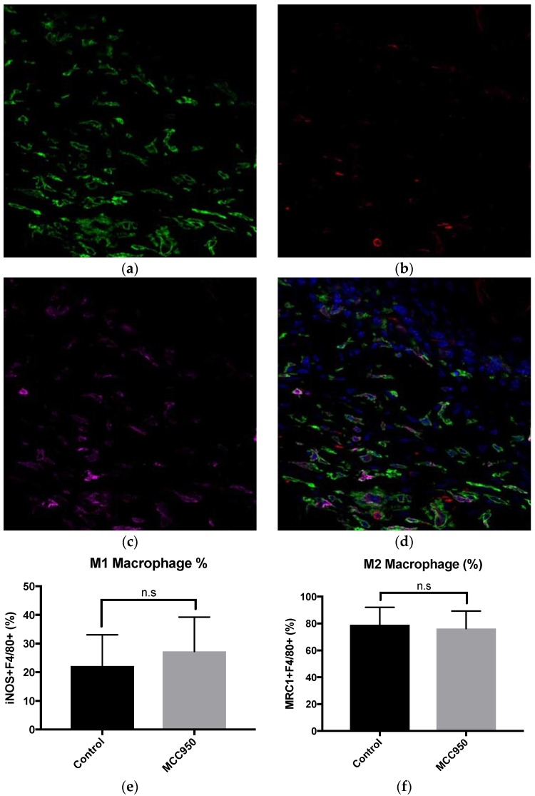
MCC950 does not alter wound associated macrophage populations. (a) Representative photomicrograph of macrophages stained with F4/80 (green). (b) iNOS to stain for M1 macrophages (red). (c) MRC1 to stain for M2 macrophages (purple). (d) A merged image of all antibodies plus DAPI for nuclear staining (blue). (e) No significant difference in M1 macrophage percentage between control and treated groups. (f) No significant difference in M2 macrophage percentage between control and treated groups. Error bars = SD. N.s. = Not significant. Scale bar = 100 μm.

