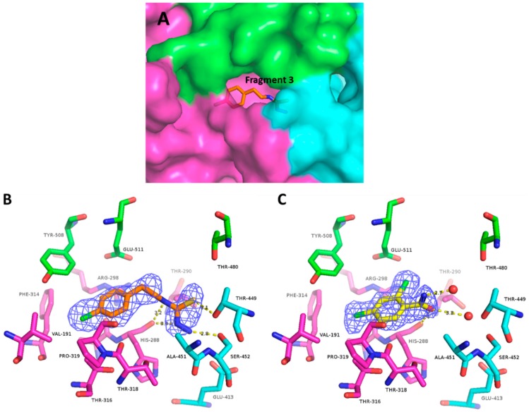
Figure 3.
Fragment-binding site B. (A) Overall view of the binding pocket with fragment 3 in place. Coloring for the three helicase domains is the same as in Figure 1. (B) Electron density map and H-bonds made by fragment 3. Residue Pro320 locates in this view on top of the fragment, and has been omitted for clarity. (C) Electron density and H-bonds made by fragment 4. Residue Pro320 has been omitted for clarity.

