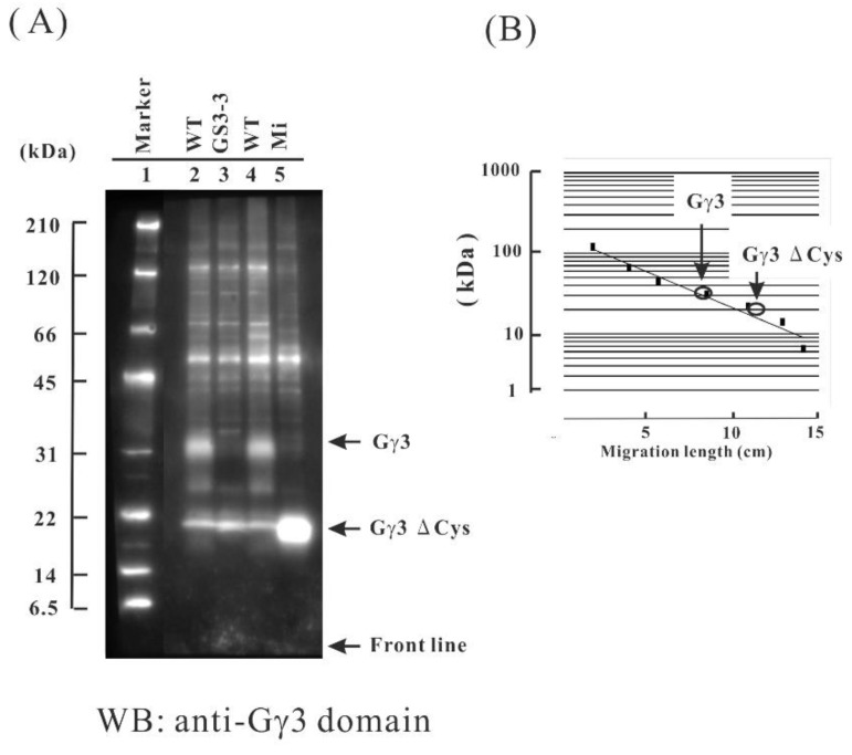
Figure 2.
Immunological study of the Gγ3 candidates in the wild-type (WT), Minute (Mi), and GS3-3 flowers. (A) First, 10 μg of each protein of the plasma membrane fractions of the WT and GS3-3 and 5 μg of the protein of the plasma membrane fractions of Mi were used for the Western blot analysis using an anti-Gγ3 domain antibody. Molecular weight marker (lane 1). The Gγ3 candidate was detected as a broad band with a molecular weight of approximately 32 kDa in the WT (lanes 2 and 4). No Gγ3 was detected in GS3-3 (lane 3). The Gγ3ΔCys candidate was detected as a band with a molecular weight of approximately 20 kDa in Mi (lane 5). (B) The molecular weights of Gγ3 and Gγ3ΔCys candidates were estimated using a molecular weight marker as a standard.

