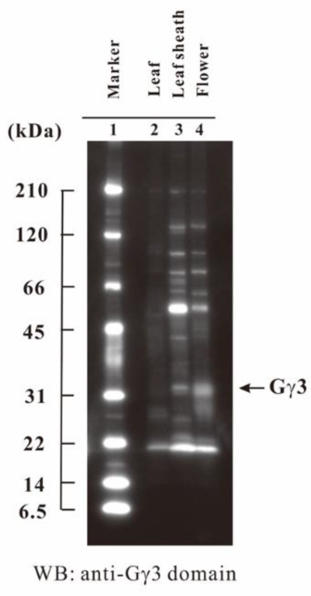
Figure 6.

Tissue-specific accumulation of Gγ3 in the wild-type (WT). Ten micrograms of each of the plasma membrane fraction proteins of the leaf, leaf sheath, and flower in the WT was analyzed by SDS-PAGE and Western blotting using an anti-Gγ3 domain antibody. Molecular weight marker (lane 1); leaf from etiolated seedling (lane 2); developing leaf sheath at the eighth leaf stage (lane 3); 1–5 cm flower (lane 4).
