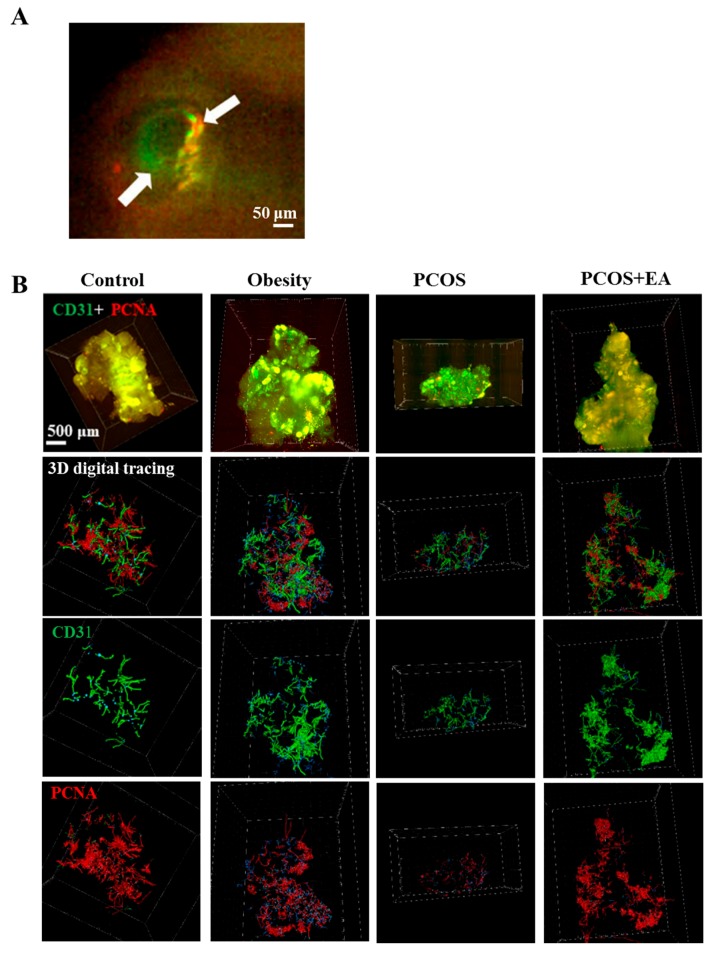
Figure 3.
EA promotes angiogenesis in PCOS-like rat ovaries. (A) CD31 was used to stain the vasculature, and proliferating cell nuclear antigen (PCNA) was used to stain the neovasculature. The white arrows indicated the co-localization of PCNA with CD31 in one follicle. (B) Ovaries from rats from different groups after CLARITY processing and immunostaining using specific antibodies, followed by data transformation using the Imaris Filament algorithm. First row: 3D rendering of whole ovary images; second row: vasculature and neovasculature within the ovaries; third row: vasculature within each ovary; fourth row: neovasculature in each ovary.

