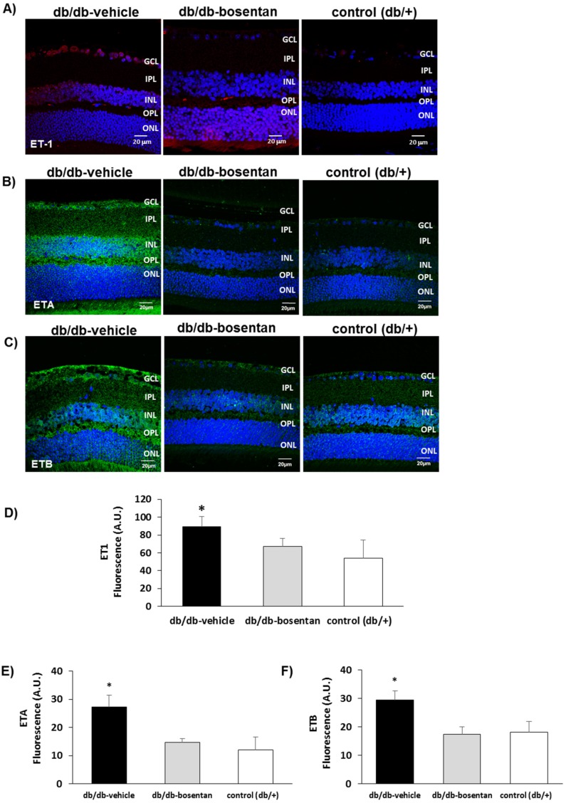
Figure 2.
(A) Comparison of endothelin-1 immunofluorescence (red) between representative samples from a diabetic mouse (db/db) mouse treated with vehicle, a diabetic mouse treated with bosentan, and a non-diabetic mouse (db/+); (B) Comparison of endothelin receptor A immunofluorescence (green) between representative samples from a diabetic mouse (db/db) mouse treated with vehicle, a diabetic mouse treated with bosentan, and a non-diabetic mouse (db/+); (C) Comparison of endothelin receptor B immunofluorescence (green) between representative samples from a diabetic mouse (db/db) treated with vehicle, a diabetic mouse treated with bosentan, and a non-diabetic mouse (db/+). GCL: Ganglion cell layer; IPL: Inner plexiform layer; INL: Inner cell layer; OPL: Outer plexiform layer; ONL: Outer nuclear layer. Scale bar: 20 µm; (D–F) Quantification of immunofluorescence. AU: Arbitrary units. Data are expressed as mean ± SD. * p < 0.05 in comparison with the other groups.

