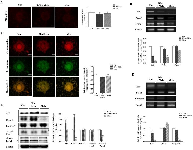
Figure 6.
Protective effects of Mito-TEMPO-like Mela response to BPA-induced ROS production, mitochondrial dysfunction, and mitochondria-mediated apoptosis in mature porcine COCs. Detection of intracellular ROS levels using DCF-DA staining in porcine COCs after BPA (75 μM) and/or Mela (0.1 μM) treatment, respectively. (A) Identification of mitochondria-specific superoxide by Mito-SOX staining in matured COCs of BPA and/or Mela treatment groups. COCs from the treated groups were stained with Mito-SOX (red fluorescence) and Mitotracker (green fluorescence) as a mitochondria detection dyes using the iRiS™ Digital Cell Image System (Korea). Scale bar = 20 µm. (B) The mRNA levels of mitochondria-related antioxidant enzymes (Sod2, Prdx3, and Prdx5) in maturing porcine COCs after BPA and/or Mela treatment were measured using RT-PCR. (C) Measurement of MMP by JC-1 staining in matured COCs after BPA and/or Mela treatment. COCs from the treated groups were stained with JC-1 to evaluate MMP (Δψm) using the iRiS™ Digital Cell Image System (Korea). Scale bar = 20 µm. (D) The mRNA levels of mitochondrial mediated apoptosis genes (Bax, Bcl-xl, and Caspase3) were investigated in BPA- and Mela-treated COCs using RT-PCR, where Gapdh was used as the internal control. (E) Western blotting results of AIF, Cyto C, Pro-Cas3, cleaved Cas3, and cleaved Parp1 in BPA- and/or Mela-treated porcine COCs as compared to the control. Relative folds of mitochondria-mediated apoptosis protein levels were obtained by normalizing the signals to β-actin. Histograms represent the values of densitometry analysis obtained using ImageJ software. Data in the bar graph are presented as the means ± SEM of three independent experiments (per 30 COCs). Differences were considered significant at * p < 0.05, ** p < 0.01, *** p < 0.001 compared to the control group (ns = not significant).

