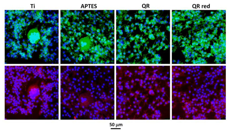
Figure 3.
Representative confocal images of multinucleated TRAP (tartrate-resistant acid phosphatase)-positive cells on the different surfaces after 5 days of culture. Cells were stained with Phalloidin-FITC (actin filaments, green), Fluoroshield-DAPI (nucleous, blue) and anti-Trap labeled with Cy3 (Trap protein, red). A higher number of osteoclast-like cells were seen on Ti and APTES surfaces.

