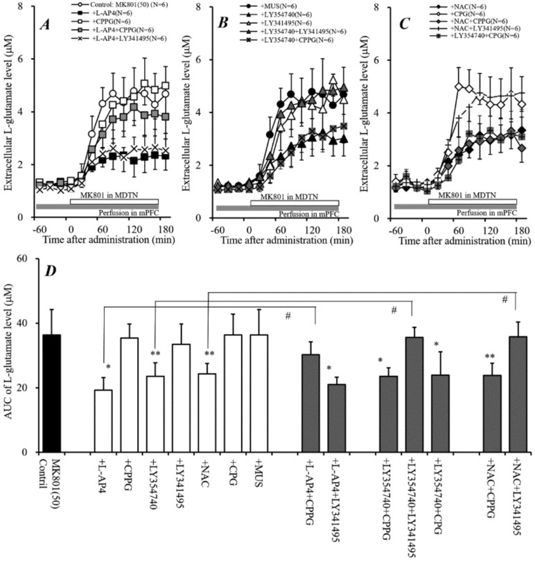
Figure 6.
Effects of local administration of modulators of Sxc, mGluRs, and GABAA receptors into the mPFC on local MK801-evoked L-glutamate release in the mPFC. (A–C) indicate the effects of perfusion with L-AP4 (100 μM), CPPG (100 μM), LY354740 (100 μM), LY341495 (1 μM), NAC (1 mM), CPG (1 mM), and MUS (1 μM) into the mPFC on local MK801-evoked L-glutamate release in the mPFC, compared with perfusion with MK801 alone from (A). Microdialysis was conducted to measure the L-glutamate release in the mPFC. In (A–C), ordinates: mean ± SD (n = 6) of the extracellular L-glutamate level in the mPFC (μM), abscissa: time after administration of MK801 (min). Open bars: perfusion of 50 μM MK801 into the MDTN. Gray bars: perfusion of 100 μM CPPG, 100 μM L-AP4, 100 μM LY354740, 1 μM LY341495, 1 mM NAC, 1 mM CPG, or 1 μM MUS into the mPFC. (D) indicates mean ± SD (n = 6) of AUC value of the extracellular L-glutamate level in the mPFC (μM) during MK801 perfusion from 0 to 180 min of (A–C). * P < 0.05, * p < 0.01; relative to Control (perfusion of 50 μM MK801 alone), and # P < 0.05; relative to perfusion of MK801 into the MDTN with L-AP4, LY354740, or NAC into the mPFC using LMM with Tukey’s post hoc test.

