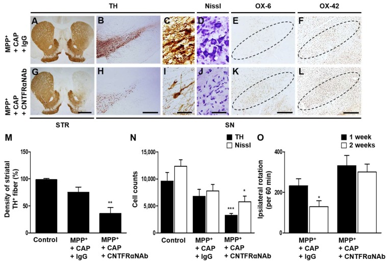
Figure 5.
Effects of CNTFRα on neuro-protection and microglial activation in the SN in vivo in MPP+-lesioned rat. MPP+ was unilaterally injected into the rat MFB followed by intranigral injection of CNTFRα neutralizing antibody (CNTFRαNAb) or IgG (control) at 1 week post MPP+ injection. All rats i.p. received CAP at 8 days post MPP+ and a continuous single injection per day for 7 days. Rats were transcardially perfused after the last amphetamine-induced rotation experiment. (A–L) Photomicrographs of TH+ fibers (A,G) in the STR, and TH+ (B,C,H,I), Nissl+ (D,J), OX-6+ (E,K) and OX-42+ (F,L) cells in the SN. (M) Optical density of striatal TH+ fibers. Student t-Test analysis, ** p < 0.01 (t = 2.720, df = 15), significantly different from MPP+ + CAP + IgG. (N) Number of TH+ or Nissl+ cells in the SN. Student t-Test analysis, * p < 0.05 (t = 1.883, df = 10), *** p < 0.001 (t = 4.355, df = 10), significantly different from MPP+ + CAP + IgG. (O) Cumulative amphetamine-induced ipsilateral rotations. Student t-Test analysis, * p < 0.05 (t = 2.239, df = 24), significantly different from 1 week. Dotted lines indicate SN. Scale bars: 1 mm (A,G), 400 μm (B,E,F,H,K,L), 40 μm (C,D,I,J). Mean ± S.E.M; (M) n = 8 to 9; (N) n = 5 to 7; (O) n = 10 to 13.

