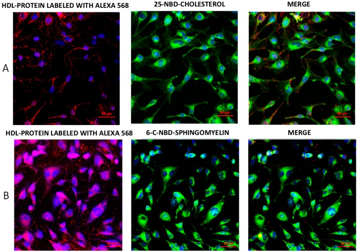
Figure 1.
Representative confocal images of lipids and high-density lipoprotein (HDL) protein internalization in HMEC-1 after 20 min incubation with fluorescent double-labeled reconstituted HDL (rHDL). (A) Cholesterol and apo AI double-labeled rHDL showed that the cellular location of protein stained with Alexa 568 (red) followed a different distribution when compared with 25-NBD-cholesterol (green). (B) Incubation of human dermal microvascular endothelial cells-1 (HMEC-1) with rHDL containing C-6-NBD-sphingomyelin and HDL protein labeled with Alexa 568 fluorescent tracers. Both sphingomyelin and protein colocalized within the cells. Nuclei were labeled with 4′,6-diamidino-2-phenylindole (DAPI) (blue). Scale bars represent 50 μm.

