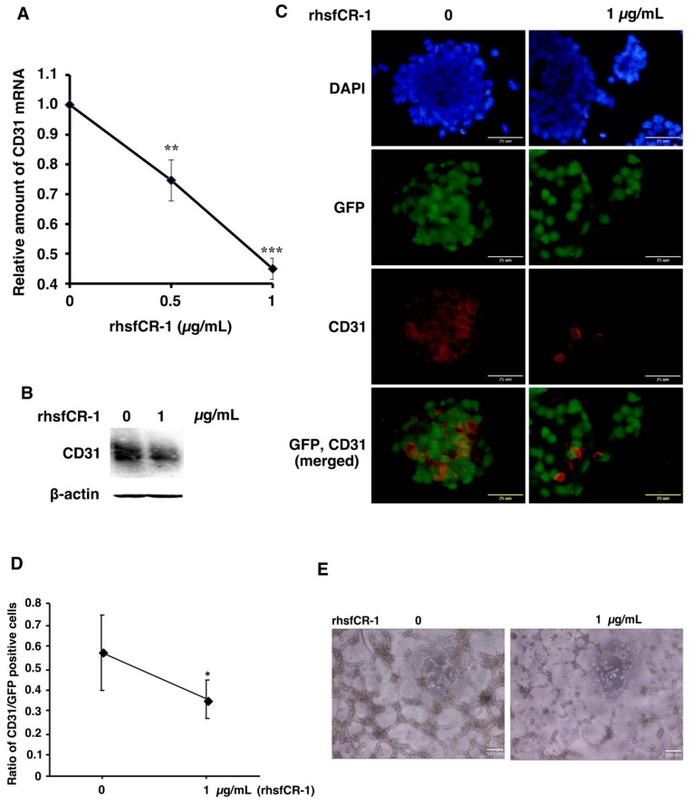
Figure 5.
rhsfCR-1 suppressed differentiation into endothelial cells using CD31+ phenotype and tube formation by miPS-LLCcm cells. (A) Relative expression of CD31 in miPS-LLCcm cells was analyzed by rt-qPCR analysis. The expression level of GAPDH was used as endogenous control. Each plot represents mean ± SD of three data points. One-way ANOVA with pairwise multiple comparison (** p < 0.01, *** p < 0.001); (B) Western blotting analysis showed the reduction of CD31 protein. Beta-actin was used as a control; (C) CD31 detected by immunofluorescence (Red) in miPS-LLCcm cells under adherent conditions. CD31 stained with anti-rabbit CD31. The nuclei were counterstained with DAPI (blue); (D) ratio of CD31-positive cells over GFP-positive cells in the absence and presence of rhsfCR-1. Student t-test was used to analyze the significance (* p < 0.05) (E) tube formation by miPS-LLCcm cells assessed in the absence or presence of rhsfCR-1 (1 µg/mL).

