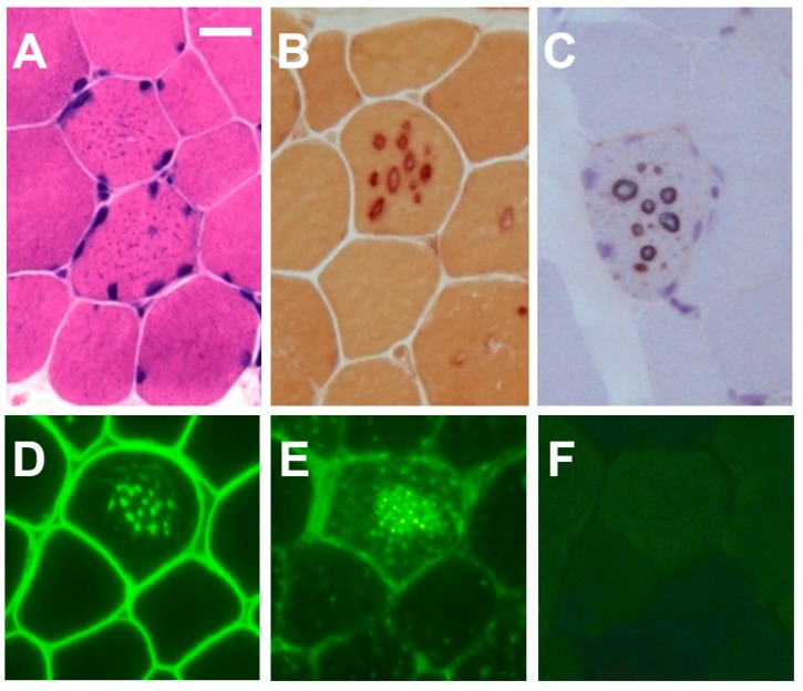
Figure 2.
Muscle pathology of a male patient with Danon disease. Tiny autophagic vacuoles look like solid basophilic granules in muscle fibers with hematoxylin and eosin stain (A). The vacuolar membrane has non-specific esterase activity (B) and acetylcholinesterase activity (C). Immunohistochemical analyses for dystrophin (D) showed that the vacuolar membrane has features of sarcolemma. Immunostaining for LIMP-1 demonstrated the overexpression of the LIMP-1 protein (E), whereas immunostaining for LAMP-2 clearly demonstrated the complete absence of the LAMP-2 protein (F). Bar = 30 μm.

