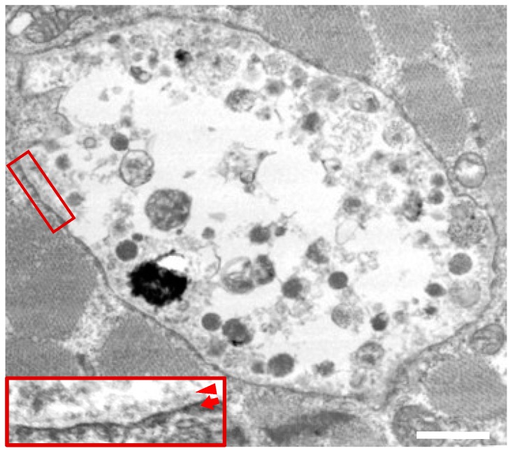
Figure 3.
Electron microscopy in skeletal muscles from a male patient with Danon disease. The vacuoles had autophagic nature as indicated by the presence of electron-dense granular materials, myeloid bodies, and variable cytoplasmic debris. In addition, basal lamina (arrowhead) are likely to be observed along the inner surface of an autophagic vacuole (arrow). The inset reveals the enlarged view of the area within the square (magnification 4×). Reconstructed with permission from our previous report [6]. Bar = 1 nm.

