Figure 2.
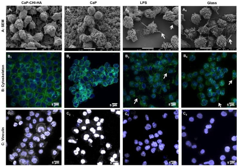
THP-1 morphology. Scanning electron microscopy (A) views after 24 h of contact with CaP-CHI-HA (A1), CaP (A2), stimulated with LPS (A3) or on glass (A4), highlighting (i) rounded cell morphology on CaP-CHI-HA and CaP (A1 and A2, respectively) and (ii) cytoplasmic extensions (arrows) on LPS and glass (A3 and A4, respectively) (scale bars indicate 10 μm). Confocal laser scanning microscopy (B) views of cytoskeleton and (C) vinculin after 24h of contact with CaP-CHI-HA (B1 and C1), CaP (B2 and C2), stimulated with LPS (B3 and C3) and on glass (B4 and C4), showing (i) concentrated F-actin at the membrane with a prominent vinculin distribution throughout the cytoplasm and the membrane in contact with CaP-CHI-HA, CaP (B1/B2 and C1/C2, respectively) and (ii) arranged F-actin in spike-like protrusions (arrows) with a peri-nuclear distributed vinculin on LPS and glass (B3/B4 and C3/C4, respectively). Green colors correspond to F-actin cytoskeleton, grey to vinculin, blue to nuclei and purple for merged vinculin/nuclei (scale bars indicate 5 μm).

